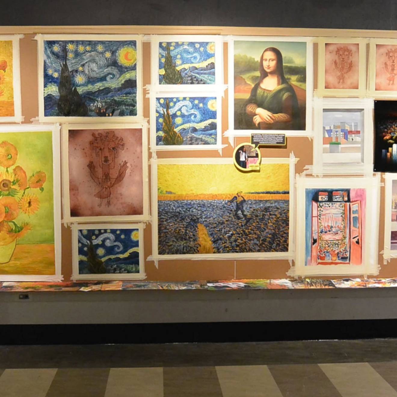
Suzanne Leigh, UCSF
For decades, doctors have noticed a rare burst of visual creativity that occurs among a small number of patients with dementia, echoing the same strange phenomenon among patients who have had a stroke or other brain injury. Now, a collaboration of 27 scientists led by UC San Francisco has offered new insights into how this talent develops as key areas of the brain degenerate.
First identified in 1996 by UCSF behavioral neurologist, Bruce Miller, M.D., visual creativity is seen occasionally in patients with frontotemporal dementia (FTD), a neurodegenerative condition affecting approximately 60,000 Americans. FTD typically affects people in their 50s and 60s, like action star Bruce Willis, whose family announced the diagnosis earlier this year. It is caused by nerve cell loss in the temporal and frontal lobes, resulting in language difficulties and behavioral changes corresponding to these two areas of the brain.
In the new study, researchers led by first author and behavioral neurologist Adit Friedberg, M.D., of the UCSF Memory and Aging Center, sought to determine the underlying science behind this unexpected artistic rebirth. Friedberg analyzed the records of 689 FTD patients and identified 17 (2.5 percent) who had experienced a sudden increase in visual artistic creativity at the start of their dementia. The study compared the brains in these newly artistic patients to those of patients who did not show increased creativity, but who shared the same FTD variant and disease stage, as well as age, sex and education level. The two groups were also matched with a third group of healthy participants.
A rise in visual creativity
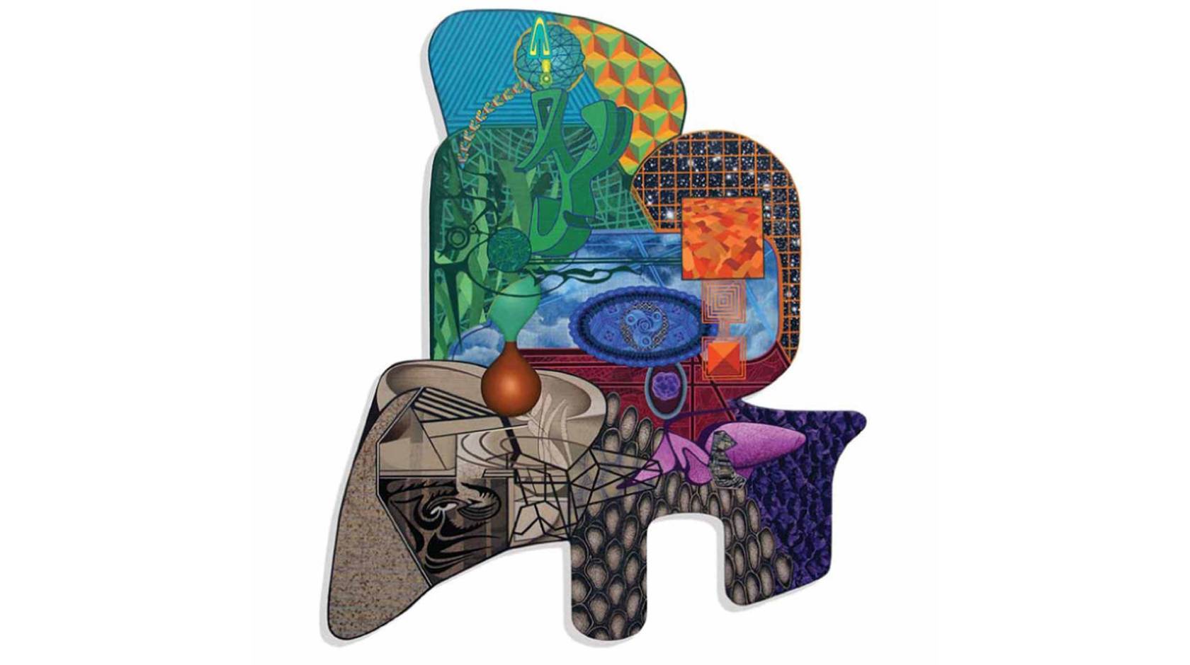
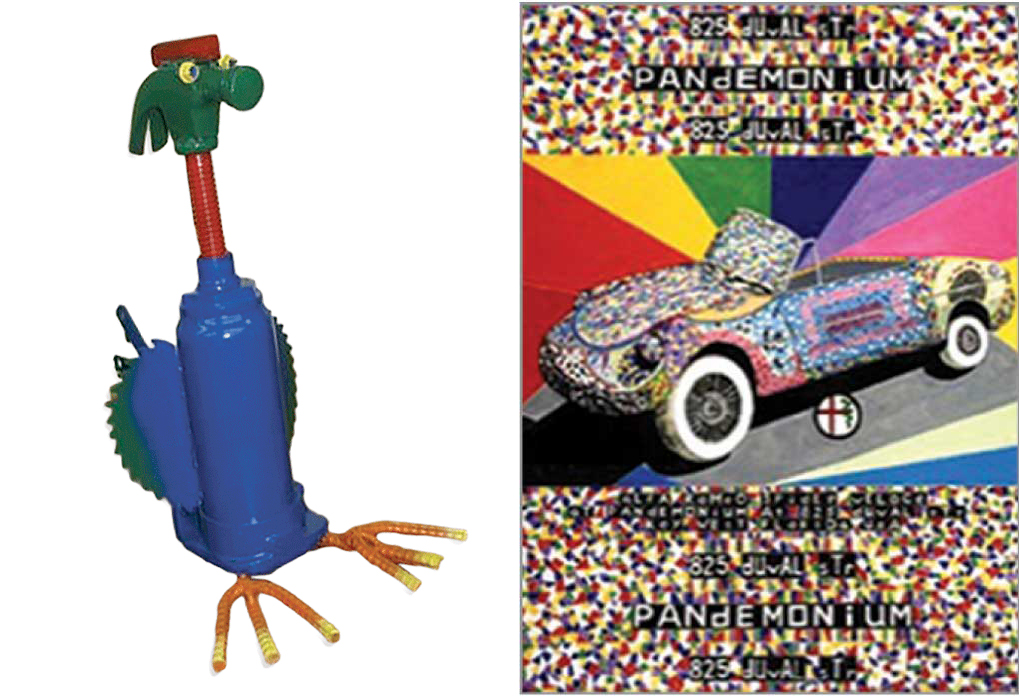
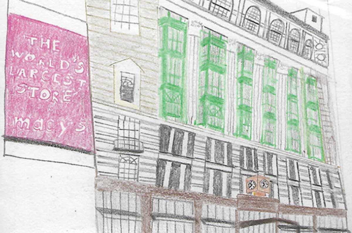
The images below are examples of artwork created by patients in the study, few of which had previous art experience. Other forms of art included painting, montage making, pottery, sculpture, jewelry making and quilting. Patients were typically engaged in their new hobby for many hours a day.
Friedberg and colleagues used neuroimaging techniques to examine multiple regions of the participants’ brains, including the frontal lobes, which are the seat of social behavior; the temporal lobes, which control the production of speech and recognition of language; and the dorsal occipital cortex, which supports visual processing.
These scans revealed that the brain regions responsible for language had shrunk in a large number of these visually creative patients, while their visual processing area showed increased activation, said Friedberg, who is affiliated with the UCSF Weill Institute for Neurosciences.
As language deteriorates, visual creativity blossoms
The results suggest that as the area of the brain controlling language deteriorates, it activates a visual processing area driving creativity, the researchers reported in their study in JAMA Neurology.
The change in brain structure was reflected in the patients’ symptoms. Among the patients with visual creativity, 10 (58.8 percent) had two variants of the disease primarily associated with difficulties understanding or pronouncing words, indicating visual creativity was more likely to occur in patients whose FTD was characterized primarily by language deficits. By contrast, only three had the behavioral form of FTD and four had variants that manifest as movement disorders.

Adit Friedberg, M.D.
“The emergence of visual creativity occurred around the onset of symptoms,” said Friedberg. “This suggests that not-yet affected brain regions may be involved and may even show increased activation.”
Eight of the 17 artists had no prior creative proclivity, the researchers found. Seven had shown some interest in visual or non-visual art and two were established artists who underwent substantial changes in their creative style. Their art included painting, montage making, pottery, sculpture, jewelry making and quilting. Artists typically engaged in their new hobby for many hours a day.
“Artwork varies from precise and mathematical to spontaneous with bright, vibrant colors and idiosyncratic content,” said Miller, the study’s senior author and director of the UCSF Memory and Aging Center. The art rarely focused on faces, but when it did, facial expressions were often bizarre, the researchers noted.
Changes in brain structure could lead to therapies
Among the study’s findings was a unique structural relationship between the brain area controlling movement in the right hand, and the visual processing area supporting creativity among the artistic patients.
Friedberg said this structural signature may reflect neuroplasticity — the ability of the brain to form new connections or reorganize itself. Understanding neuroplastic processes in this context may add to the arsenal of strategies to combat FTD and other neurodegenerative disorders, she said.
It is not known why some patients develop emergent creativity, but genes are likely to play a role, Miller said.
“Our research shows that this cohort was more likely to perform better on a cognitive test than their non-artistic counterparts,” he said. “It may also be likely that these individuals operate in an environment conducive to creativity.”
Co-authors, Funding and Disclosures: The study involved a collaboration between 24 researchers in neurology, radiology and pathology at UCSF, as well as colleagues in neurology, genetics and precision medicine from UCLA and three institutions in Ireland and Barcelona. For details, please see the paper.
